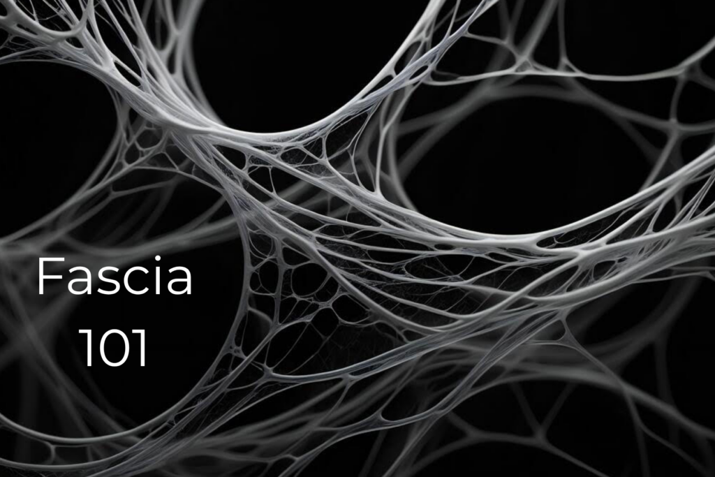
The most important thing to keep in mind is that fascia is living tissue—and it is one continuous structure throughout the entire body. Rather than existing as separate pieces, the fascial network forms an interconnected system that surrounds, supports, and connects every muscle, bone, nerve, blood vessel, and organ.
To make this easier to understand, fascia is commonly divided into four categories based on its location and function within the body.
1. Superficial Fascia
Superficial fascia is often described as a layer of loose connective tissue located just beneath the skin and the slightly more superficial layer of adipose tissue (body fat).
Although fibrous, it is also highly elastic. It separates the skin from the muscles, allowing the two to glide smoothly over one another during movement. This layer also plays an important role in:
- Thermoregulation (body temperature control)
- Circulation
- Lymphatic flow
- Connecting with the deeper fascial layers
2. Deep Fascia
Deep fascia is a dense, highly organized fibrous layer that surrounds and covers the muscles. Imagine it as a full-body “catsuit” or body stocking that holds everything together.
This is the layer where myofascial force transmission occurs.
For example, when you simply pick up a cup of coffee or tea, an intricate sequence of force is transmitted through your shoulder, elbow, wrist, and fingers. The fascia is actively involved throughout this entire movement, helping transmit those forces efficiently throughout the body.
Research suggests that as much as 30% of muscular tension may be transmitted through fascial tissues rather than muscle alone.
3. Meningeal Fascia
The meningeal fascia surrounds the brain and nervous system.
This topic alone could fill multiple chapters, but from both a movement and mind-body perspective, one concept is especially important:
The nerves responsible for proprioception—your body’s awareness of where it is in space—are sensory nerves embedded within the fascia.
In fact, fascia is considered the body’s largest sensory organ.
Within a typical muscle, there are approximately three times as many sensory neurons as motor neurons. This highlights an important principle: the body’s need for sensory awareness and refinement is actually greater than its need for motor control.
Research using hypertonic saline injections demonstrated that the thoracolumbar fascia is more sensitive to pain than the underlying erector spinae muscles. Participants most commonly described fascial pain as:
- Burning
- Throbbing
- Stinging
Overall, researchers concluded that fascia is one of the most pain-sensitive tissues of the lower back.
4. Visceral Fascia
Visceral fascia surrounds the body’s internal organs, including the lungs, heart, and abdominal organs.
It suspends these organs within their cavities while allowing the normal movement necessary for healthy physiological function. This fascial system includes visceral ligaments that both anchor the organs to the body wall and permit the motion required during breathing, digestion, and everyday movement.
The Origin of Biomechanics
In 1680, Giovanni Alfonso Borelli, often referred to as the father of biomechanics, compared the bodies of humans and animals to machines functioning through an intricate system of beams, pulleys, and levers.
Today, this concept has evolved into what is known as biotensegrity.
Rather than viewing the skeleton as a rigid framework supporting the body, biotensegrity proposes that the body’s 206 bones function as compression elements suspended within a continuous tension network created by fascia, ligaments, and tendons. Together, these tissues help maintain posture, absorb force, and resist gravity.
As the founder of osteopathic medicine, Andrew Taylor Still wrote in 1889:
“There is no real difference between structure and function; they are two sides of the same coin. If structure does not tell us something about function, it means we have not looked at it correctly.”
That principle continues to guide modern fascial assessment today.
Understanding Fascial Conditions
Before treatment begins, a Pathoanatomical Analysis (PAA) is performed along with a detailed postural assessment.
In simple terms, four primary postural distortions are evaluated:
- Shifts
- Tilts
- Bends
- Rotations
Many people present with a combination of these patterns rather than just one.
Once the assessment is complete, additional diagnostic tools may be used when appropriate.
For example, ultrasound imaging has shown that the posterior layer of the thoracolumbar fascia is, on average, 25% thicker in individuals with chronic low back pain compared with those who do not experience back pain.
Another valuable tool is ultrasound elastography, which combines traditional ultrasound imaging with the ability to measure tissue stiffness while producing color-coded images of tissue elasticity.
The concept of tissue elasticity traces back to Robert Hooke, the English scientist who first described the relationship between force and elasticity in 1660.
Understanding both the structure and mechanical properties of fascia provides valuable insight into pain, movement dysfunction, and recovery.
How Does Pilates Help Fascia?
In Part II, we’ll explore:
- The role of connective tissue in pain and mental health
- How fascia shapes movement
- Why healthy fascia reduces stiffness
- Improving mobility and flexibility
- Easier, more efficient breathing
- Better posture
- Enhanced body awareness and movement efficiency
- How Pilates helps restore healthy fascial function throughout the body
Stay tuned as we dive deeper into the remarkable world of fascia and why caring for this living tissue can transform the way you move and feel.